


A guest post by Jaden Graham
I have the pleasure of teaching our Speaking Your Truth: Content Creation for Birth Workers course once a year for the Indie Birth Midwifery School, and I love sharing (with permission!) some of our standout pieces that students create. It is so exciting to see awesome content generated, and to think about the tangible ways this will positively impact our birthing world. This piece taught me a lot, and was fascinating to geek out on. Enjoy!
—————————————————–
The umbilical cord is the lifeline that connects the growing baby to the placenta in utero. Developing around the fifth week of pregnancy, the umbilical cord is a sinewy vessel that carries nutrients and oxygenated blood to the fetus (via the umbilical vein) and removes carbon dioxide and other waste material from fetus to placenta (via the umbilical arteries) to maternal blood circulation where they can be processed and excreted – acting as both a nourishment and filtration conduit on behalf of the placenta. In about 90% of pregnancies, the cord implants itself into the center of the placenta, commonly known as a “normal” cord insertion; however, sometimes, “normal” can present itself as the cord attaching to the edge of the placenta (known as a marginal insertions), as well as inserting into the placental membranes away from the edge of the placenta or extending across like tree roots between the amnion (inner membrane surrounding the baby; the amniotic sac) and chorion (outer membrane) before reaching the placenta, called a velamentous cord insertion. I want to emphasize that these conditions, while not necessarily deemed by the medical industrial complex as “normal”, they are, nonetheless, common, and also while this anomaly can develop into high risk scenarios requiring immediate medical attention, that abnormal cord insertions, specifically velamentous cord insertions (VCI), they should not automatically result in a cesarean section.
Velamentous cord insertions occur in about 1.1% of singleton pregnancies and 8.7% in twin gestations, having a higher occurrence in monochorionic placentation of identical twins when the placentas are fused into one versus when the placentas are non-fused.4 Marginal cord insertions occur in about 7% in singleton pregnancies and 25% of twin pregnancies5, respectively, and have the potential to develop into a velamentous cord insertion as pregnancy progresses. For the remainder of this article, I will focus on the latter, and more complex, condition, velamentous cord insertions.
There are currently two theories on why velamentous cord insertions occur. One theory holds that when the blastocyst (the precursor to the embryo and fetus) attaches itself to the uterus in early pregnancy, the border of the yolk sac (the source of nutrition to the growing embryo before the formation of the placenta, also known as a fetal pole) is oriented away from the endometrium as opposed to when it is commonly positioned towards it. As a result, insertion of the cord materializes in what ultimately becomes the chorionic laeve, the outer layer of the placenta.6 The other theory proposes that the placenta may go through a process known as trophotropism. Like a plant leaning towards the sun throughout a given day to acquire the sunlight it needs to survive, a placenta that seeks the best blood supply can grow in the direct of favorable blood supply and can atrophy where the blood supply is less favorable.”7 Higher uterine blood supply is more favorable than in the lower uterine segment and sometimes, if the placenta implants low, it can essentially migrate to achieve greater blood supply; however, in its migration process, every other existing part of the placenta, including the cord, remains where it formed.7
Concurrent with the presence of VCI is the absence of Wharton’s Jelly. “Wharton’s Jelly is gelatinous substance that provides insulation and protection within the umbilical cord.”8 In the case of VCI, since the blood vessels travel across between the amnion and chorion before reaching the placenta, they are, therefore, unprotected by Wharton’s Jelly where they cross the placental membranes before coming together to form the umbilical cord.9
As previously stated, a VCI can, in fact, create certain high-risk scenarios (albeit, rarely). The most notable complication is a condition known as vasa previa, which occurs in 6% of pregnancies with a VCI4 or in 1:2000-3000 of all pregnancies7; although, this figure has been reported to vary from 1:513-6000 in naturally conceived pregnancies and 1:293 in IVF-treated pregnancies10 (interestingly, on the same website). Vasa previa is a placental anomaly in which exposed fetal vessel lie too close or on top of the cervical os (opening) and in front of the baby’s presenting part. The risk lies in the vessels tearing during active labor as the cervix dilates, membranes rupturing (especially in the case of PROM), or the vessels becoming “pinched off as they are compressed between the baby and the walls of the birth canal”10 – all of which can lead to severe fetal blood loss and potentially death. The infant mortality rate of an undiagnosed vasa previa is 50-95%.10
While vasa previa is indeed a rare condition, we cannot bypass the fact that when undiagnosed it does carry significant risk, and it is not the intention of this paper to suggest otherwise; however, it is very important to note that vasa previa, in conjunction with a VCI, presents a risk only when the placenta is located in the lower uterine segment, not high lying. According to the International Vasa Previa Foundation, a VCI in the upper segment of the uterus is rarely of great consequence: “When located in the upper segment of the uterus the risks are not as great as when located in the lower segment.”7 The writer goes on to state that there is a chance that the blood vessels have the potential to rupture during third stage of labor if the cord is pulled; although, other sources, have noted that this is rare and that with the absence of vasa previa, complications are also rare. Still, care should be taken with the delivery of the cord to minimize the risk of the cord tearing and to avoid excessive traction prior to placental separation.11
Unfortunately, despite the distinction between high and low lying placentas and the risks they carry, certain popular “family health” websites, such as Very Well Family, explain VCIs in “blanket medicine” terms where it is automatically a “complication” and goes on to say, “If you have been diagnosed, your doctor may want to perform a c-section,”12 without acknowledging the aforementioned distinction.
Also, on many medical studies of VCI and postpartum outcomes the associated, observed “risks” of VCI are 5 min low Apgar scores (below 7), low birth weight, increased NICU admissions, and increase in cesarean.13 I want to critique here a few of the above factors that could’ve led to these adverse outcomes.
It has been found that prenatally, a VCI can restrict fetal growth.14 While I have not found any articles that explain exactly why, I can only hypothesize that because of inserting on the outside of the placental disk via fetal membranes versus in the common central insertion site – essentially due to this extra step a VCI has to go through to reach the placenta – that nutrient uptake may not be as well achieved as it would in a baby with a normal cord insertion. If this is the case, and vasa previa is not present, wouldn’t you want to have the pregnant person continue gestation for as long as their bodies want them to in order to allow the baby to grow to a “desirable” weight? Apparently, this is not the case as in one study, which specifically evaluated the perinatal outcomes of singleton and twin pregnancies without concurrent vasa previa, one of the primary outcomes included cesarean or surgical vaginal delivery “for a nonreassuring fetal heart rate pattern.”13 The mean gestational age was only 36.2 weeks – not incredibly early in pregnancy but quite early for a baby that might have growth restriction due to a VCI. These two outcomes paralleling each other only begs the question: are these parents electing to have a cesarean via their doctors advice or are their bodies going into labor on their own divine accord? And if the former, is it even appropriate to force the process of birth for a baby with stunted intrauterine growth? And also, because this process may very well be forced and these babies, in fact, don’t, have the sufficient amount of nutrition built up in their bodies that allowing time and continued growth would have given them, is this a direct correlation to the adverse incidence of NICU admission, low Apgar scores of below 7 (after 5 minutes), and even, in some circumstances, neonatal death?
In conclusion, there are still many questions that need to be answered and many holes in the bigger picture of velamentous cord insertion, as apparent “risks” that VCIs carry may instead be the misunderstanding of this diagnosis by doctors. Also, in cases and further research of VCI, care should be taken to assess whether or not the placenta is located in the upper or lower uterine segment to ultimately determine if this variance makes a difference in the health, safety, and delivery of both baby and birthing person. I personally believe that in assessing perinatal outcomes and the “true” risk they carry, it is absolutely critical to make this distinction – in both research studies and simple clinical settings- to accurately “measure” said risks. It is also in my personal experience with placentas located in the upper uterine segment that the best course of action is inaction, which no hard data has revealed.
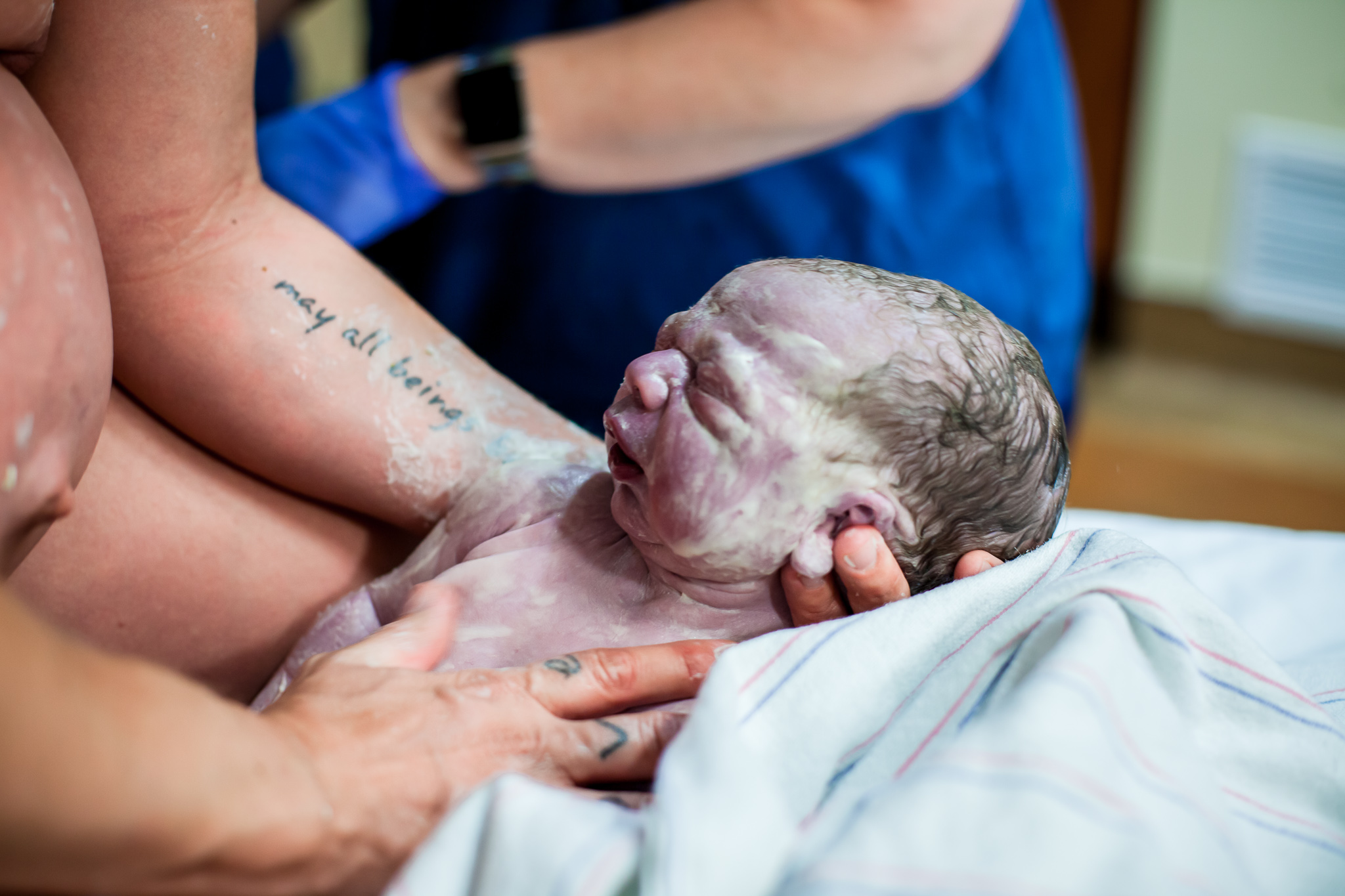
At 43 weeks and 6 days of my pregnancy, after many failed attempts at self-induction and a PROM, my midwife intuitively advised and arranged for my transfer to the hospital where I was induced via Pitocin. I went into active labor 6 hours after being admitted and 4.5 hours later, my son was born – 6 lbs. 13 oz. and a surprise velamentous cord insertion. His Apgar score was 9 after 1 minute. I found out months later that had an ultrasound diagnosed the VCI when I was pregnant, that the routine of the hospital I had received this at was to put the patient on bed rest for the remainder of their pregnancy and to have an elective cesarean at 35 weeks. How grateful I am that I never found out and, in reflection, the best thing for him was to take those extra weeks to obtain the growth that he so needed – my body and my baby both very wise and in synchronicity with each other.
So let me reiterate, velamentous cord insertions alone should not automatically result in a cesarean section. If anything, when no risk other than its existence presents itself, it seems that it is best that we be patient with our bodies, our babies, and ourselves, and allow them the time they need to grow and be nourished.
Jaden Graham is an Indie Birth Midwifery School student, Western folk herbalist, and full spectrum doula serving pregnant persons and families in Northern California. You can find more of her magic, medicine, and musings on Instagram @thankful_earth and @spiralpathbirth.
References
1. Ebbing, Cathrine et al. “Prevalence, risk factors and outcomes of velamentous and marginal cord insertions: a population-based study of 634,741 pregnancies.” PloS one vol. 8,7 e70380. 30 Jul. 2013, doi:10.1371/journal.pone.0070380
2. Spurway, Jacqueline et al. “The development, structure and blood flow within the umbilical cord with particular reference to the venous system.” Australasian journal of ultrasound in medicine vol. 15,3 (2012): 97-102. doi:10.1002/j.2205-0140.2012.tb00013.x
3. Schöni‐Affolter F, Dubuis‐Grieder C, Strauch E. The umbilical cord. [Internet] 2007 [updated 15th October 2007; cited 2012 10th March]; Available from: http://www.embryology.ch/anglais/fplacenta/cordon01.html.
4. Velamentous Cord Insertion. International Vasa Previa Foundation. http://vasaprevia.com/Velamentous-Cord-Insertion. Retrieved on September 9, 2019.
5. Skalina, Tristan & Weerakkody, Dr. Yurunga et al. Marginal Cord Insertion. Radiopaedia. https://radiopaedia.org/articles/marginal-cord-insertion/ Retrieved on September 9, 2019.
6. Curtin, WM et al. “Accuracy of fetal anatomy survey in the diagnosis of velamentous cord insertion: a case study control.” Dovepress. https://doi.org/10.2147/IJWH.S189718 Published 11 March 2019. Volume 2019:11. Pg. 169-176.
7. Frequently Asked Questions. International Vasa Previa Foundation. http://vasaprevia.com/Frequently-Asked-Questions Retrieved on September 9, 2019.
8. Shiel Jr. MD, William C. “Medical Definition of Wharton’s Jelly.” MedicineNet. https://www.medicinenet.com/script/main/art.asp?articlekey=24387 Retrieved on September 10, 2019.
9. Hill, M.A. Embryology Placenta – Abnormalities. Retrieved from https://embryology.med.unsw.edu.au/embryology/index.php/Placenta_-_Abnormalities. Retrieved on September 10, 2019
10. Vasa Previa Defined. International Vasa Previa Foundation. http://vasaprevia.com/Vasa-Previa-Defined Retrieved on September 11, 2019.
11. Oyelese, Yinka. “Obstetrics and Gynecology: Velamentous Cord Insertion.” Cancer Therapy Advisor. https://www.cancertherapyadvisor.com/home/decision-support-in-medicine/obstetrics-and-gynecology/velamentous-insertion/ Retrieved on September 11, 2019.
12. Danielsson, Krissi. “Velamentous Cord Insertion Pregnancy Complications.” Very Well Family. https://www.verywellfamily.com/velamentous-cord-2371665 Updated June 15, 2019.
13. Sinkin MD, Joshua A. et al. “Perinatal Outcomes Associated With Isolated Velamentous Cord Insertion in Singleton and Twin Pregnancies” Journal of Ultrasound Medicine. Volume 37. 2017. Pg. 471-478.
14. Ebbing, Catherine et al. “Velamentous or marginal cord insertion and the risk of spontaneous preterm birth, prelabor rupture of the membranes, and anomalous cord length, a population‐based study.” Acta Obstetricia et Gynocologica Scandanavia. Vol 96, Issue 1. October 3, 2016. Pg. 78-85.


Quite the detailed descriptions of cord insertions. I am very glad you and your baby were not diagnosed to have a cesarean section. Thanks for the research and the call to inaction in a VCI.
Amazing paper! Thank you for the information. May I share?
Yes, please share!
Very happy your baby was born healthy, with no complications! When I first read this article, it made me feel a little more calm and I loved how the 2 theories were pointed out regarding possible reasons the VCI happens in some pregnancies. However, I just can’t ignore that yes, there are still black holes and unanswered questions, regarding the outcome of natural birth with a VCI condition. The risks are still there for things to go really bad with versa previa or not, with placenta at upper segment or lower segment, and regardless of the actual location of insertion, all possibilities are still there! Anything can happen, even a rupture of vessels at any given moment. This gives me so much anguish and worry. I continue to lean towards a c-section. Natural birth is beautiful but in this situation the price is too high, and I don’t want to take chances.
Thank you for writing this detailed article, it’s helping me to navigate how to approach my experience. I’m so grateful for a more nuanced perspective.